• This website has been created and funded by AstraZeneca UK for educational purposes only and is intended for UK Healthcare Professionals only. It should not be used for diagnosing or treating a health problem or disease.
  • Stay informed


SEE THE PATTRNS Logo
  • Home
  • About ATTR amyloidosis
    • Overview
    • Signs and symptoms
    • Diagnosis
  • Phenotyping HF
  • Referral
  • Management approaches
  • Resources

PHENOTYPING HEART FAILURE (HF)

PHENOTYPING HF CAN SUPPORT ACCURATE DIAGNOSIS AND HELP FACILITATE THE TIMELY INITIATION OF TREATMENT1–4gradient-line

What is phenotyping in HF?

divider line

Phenotyping is the process of characterising HF beyond ejection fraction (EF), using clinical features, biomarkers, and imaging to help identify underlying causes of HF.5 This deeper understanding provides a clearer view of the pathophysiological mechanisms driving HF in each individual patient.6,7
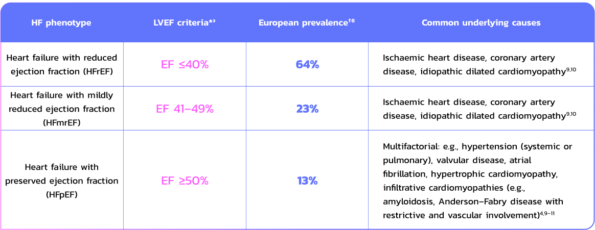
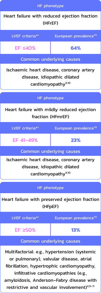
HF phenotypes differ in their clinical features and underlying causes:3

table table

*Based on the 2021 ESC Guidelines for the diagnosis and treatment of acute and chronic HF;2

†Derived from a prospective, observational study of 9,134 patients with HF in the ESC Heart Failure Long-Term Registry. Countries included Lithuania and Sweden (northern countries); Bosnia and Herzegovina, Bulgaria, the Czech Republic, Hungary, Latvia, Poland, Romania and Slovakia (eastern countries); Austria and France (western countries); Greece, Italy, Portugal, Serbia, Slovenia, Spain and Turkey (southern countries); Israel (Middle East), and Egypt (North Africa).8

From measurement to mechanism: EF shows cardiac function, phenotyping can help reveal the cause

divider line

Why is phenotyping important?

  • HF is not a single condition – symptoms, causes, and progression can vary widely between phenotypes2,7
  • Managing HF symptomatically, without investigating underlying causes, may lead to missed opportunities for targeted, disease-specific therapy5,11,12
  • By identifying the root cause of HF, phenotyping moves away from a “one size fits all approach”5 and enables more precise and personalised management, as well as facilitating timely treatment and earlier intervention1,2,12

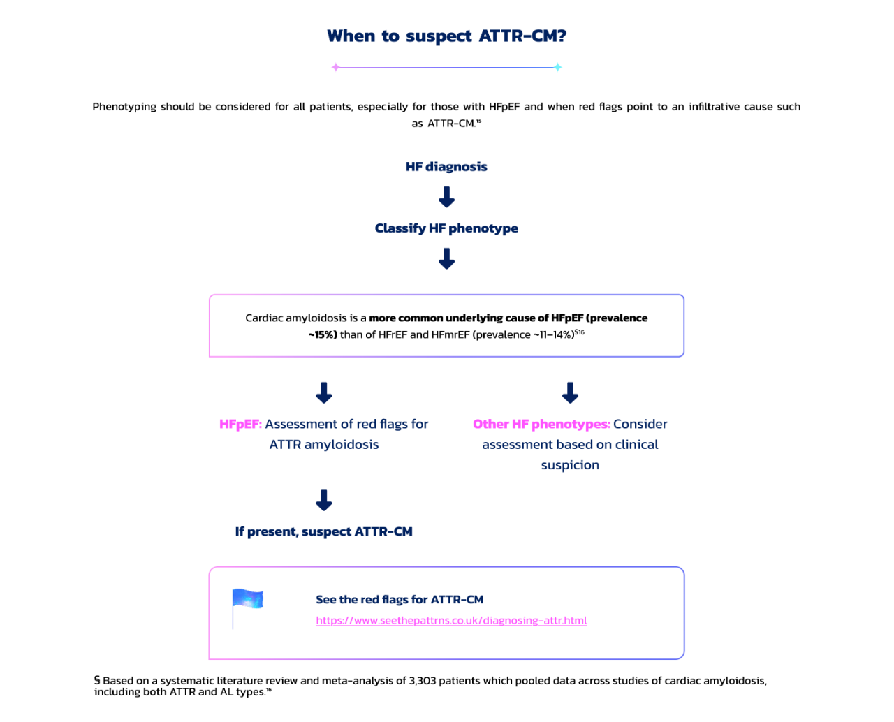
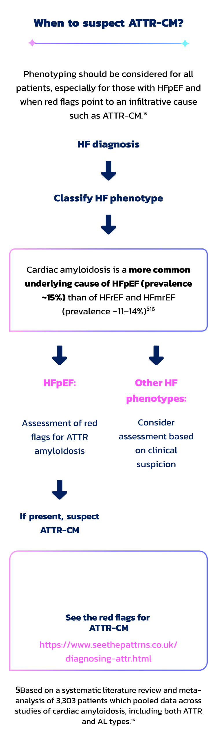
For example, in high-risk patients with HFpEF or HFmrEF aged ≥60 years, the prevalence of transthyretin-mediated amyloidosis with cardiomyopathy (ATTR-CM) in Europe may be as high as 24%‡13

In patients with HF due to ATTR amyloidosis, median survival can be as short as 2.6 years from diagnosis14

heart-beat

As such, early disease identification, through phenotyping HF, can help improve patient outcomes1,4

‡Europe: 24% (95% CI: 18.2–30.2), comprised of Italy: 41% (95% CI: 23.7–59.4; 13/32); United Kingdom: 40% (95% CI: 19.1–64.0; 8/20); France: 29% (95% CI: 10.3–56.0; 5/17); Spain: 20% (95% CI: 13.1–29.0; 22/109); Poland: 6% (95% CI: 0.8–20.8; 2/32). "High risk" criteria included age ≥60 years with a history of HF, LVEF >40%, an end-diastolic interventricular septum thickness ≥12 mm, but without diagnosed amyloidosis, history of LVEF ≤40%, cardiomyopathy of known cause, severe valvular, or coronary heart disease.13

suspect suspect

What to do if ATTR-CM is suspected?

divider line
  • To rule out AL amyloidosis (which requires different treatment approaches to ATTR-CM): Perform assessment with serum-free light chain quantification and urine immunofixation17
  • To confirm ATTR-CM:
    • Cardiac magnetic resonance imaging (CMR) to identify features suggestive of amyloidosis, such as restrictive morphology and abnormal T1 mapping17
    • Bone scintigraphy (99mTc-PYP or DPD scan) to support diagnosis in absence of a monoclonal protein17

See the ATTR-CM diagnostic pathway

https://www.seethepattrns.co.uk/diagnosing-attr.html

Integrating phenotyping into routine clinical assessments may help uncover underlying causes of HF and guide timely, effective treatment1–4

divider line

A multi-modal approach to HFpEF phenotyping may improve diagnostic accuracy of underlying conditions such as ATTR-CM:5,6,18,19

Clinical features2,19

  • Symptoms and signs
  • Comorbidities
  • Physical exam findings (e.g., blood pressure, heart rate, evidence of congestion)

Biomarkers2,5,19

  • Natriuretic peptides (BNP and NT-proBNP)
  • Cardiac injury markers (e.g., high-sensitivity cardiac troponins)
  • Markers of fibrosis (e.g., soluble ST2)

Imaging2,19

  • Echocardiography
  • Cardiac magnetic resonance imaging (CMR)
  • Nuclear imaging (e.g., bone scintigraphy)

Early suspicion of ATTR amyloidosis and subsequent referral by HF-specialist cardiologists can support accurate diagnosis and appropriate treatment1,2

UNDERSTAND REFERRAL


99mTc-PYP=technetium-99m-pyrophosphate; AL=light chain amyloidosis; ATTR=transthyretin amyloidosis; ATTR-CM=transthyretin amyloidosis with cardiomyopathy; BNP=B-type natriuretic peptide; CI=confidence interval; CMR=cardiac magnetic resonance imaging; HF=heart failure; HFmrEF=heart failure with mildly reduced ejection fraction; HFpEF=heart failure with preserved ejection fraction; HFrEF=heart failure with reduced ejection fraction; NT-proBNP=N-terminal pro-B-type natriuretic peptide; ST2=suppression of tumorigenicity 2.


References: 1. Jasinska-Piadlo A and Campbell P. Heart. 2023;109(11):874–883; 2. McDonagh TA, et al. Eur Heart J. 2021;42(36):3599–3726; 3. Bonfioli GB, et al. Eur Heart J Suppl. 2025;27(Suppl 1):i115–i121; 4. Madan N and Kalra D. Rev Cardiovasc Med. 2020;21(2):181–190; 5. Senthong V, et al. Curr Heart Fail Rep. 2017;14(2):106–116; 6.  Shah SJ, et al. Heart Fail Clin. 2014;10(3):407–418; 7. Roh J, et al. Circ Res. 2022;130(12):1906–1925; 8. Chioncel O, et al. Eur J Heart Fail. 2017;19(12):1574–1585; 9. Zhou Q, et al. Front Cardiovasc Med. 2021;8:678121; 10. Shang Z, et al. Rev Cardiovasc Med. 2022;23(1):030; 11. Zawadzka MM, et al. Adv Clin Exp Med. 2023;31(10):1163–1172; 12. Kwok CS, et al. J Cardiovasc Dev Dis. 2022;9(12):455; 13. Yun S, et al. Amyloid. 2024;31(4):291–301; 14. Dungu JN, et al. Circ Heart Fail. 2016;9(9):e003352; 15. Anker SD, et al. Eur J Heart Fail. 2023;25(7):936–955; 16. See ASY, et al. Heart Lung Circ. 2022;31(11):1450–1462; 17. Gillmore JD, et al. Circulation. 2016;133(24):2404–12; 18. Alwan L, et al. JACC Cardiovasc Imaging. 2024;17(2):195–211; 19. Kim IC and Yoo BS. Diagnostics (Basel). 2022;12(6):1366.

GB-69505 | November 2025

WELCOME TO THE SEE THE PATTRNS WEBSITE

This is a non-promotional AstraZeneca website intended for UK healthcare professionals.

By clicking "I am a UK Healthcare Professional" you are declaring and confirming that you are a Healthcare Professional within the UK and have read and understood this disclaimer. Clicking "I am NOT a UK healthcare professional" will take you to www.astrazeneca.co.uk

Patients or members of the public: This website is intended for healthcare professionals only. Please speak to your healthcare provider if you require further information about ATTR amyloidosis.

I AM NOT A UK HEALTHCARE PROFESSIONAL
  • AstraZeneca Logo
  • Cookies
  • Legal Notice and Terms of Use
  • Privacy Notice
  • Contact us

The information on this website has been created and funded by AstraZeneca UK for educational purposes only. The content is intended for UK healthcare professionals and should not be used for diagnosing or treating a health problem or disease.


©2025 AstraZeneca. All rights reserved. GB-70859 | December 2025